OCT Angiography: A Revolutionary Tool for Eye Health
Optical Coherence Tomography Angiography (OCT-A) is an advanced imaging technology that is changing the way doctors diagnose and treat eye diseases. It’s a non-invasive procedure that allows doctors to see blood vessels in the eye in great detail without needing to inject dye into the bloodstream. OCT-A has become especially important for managing conditions like macular degeneration, diabetic retinopathy, and glaucoma. Let’s take a closer look at what OCT-A is and the many benefits it offers for eye health.
What is OCT Angiography?
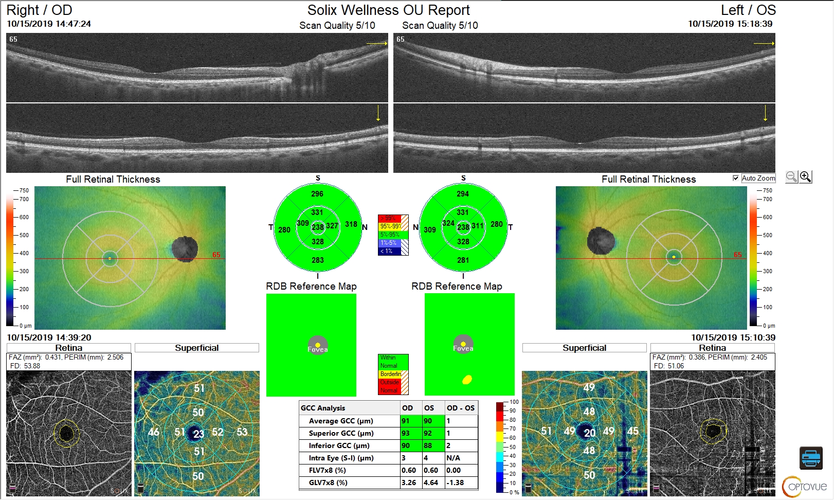
OCT Angiography uses light waves to take pictures of the inside of the eye. It works similarly to traditional OCT, which uses light to create detailed cross-sectional images of the eye’s layers. However, OCT-A goes one step further by focusing on the blood vessels in the retina. It uses changes in the light reflection caused by the movement of blood cells to create a detailed map of the blood flow in the eye.
The retina, located at the back of the eye, is where vision begins. It contains tiny blood vessels that provide oxygen and nutrients to the eye. Problems with these blood vessels can lead to serious eye conditions that threaten vision. OCT-A helps doctors spot these problems early, before they cause permanent damage.
How Does OCT Angiography Work?
OCT Angiography works by using a special device that sends light waves into the eye. These waves bounce back off the retina, and the device measures how long it takes for the light to return. By looking at how the light reflects off moving blood cells, the machine creates images that show blood flow within the eye.
Unlike older techniques, OCT-A does not require any dyes or injections. Traditional methods of eye imaging, like fluorescein angiography, need a dye to be injected into the bloodstream, which can cause discomfort or even allergic reactions. OCT-A eliminates this step, making it safer and more comfortable for patients.
Benefits of OCT Angiography
Non-invasive: One of the biggest advantages of OCT-A is that it doesn’t require needles or injections. Traditional methods, like fluorescein angiography, involve injecting dye into the veins to highlight the blood vessels. This can be uncomfortable and carries a risk of side effects. OCT-A is pain-free and much safer.
Early Detection: OCT-A allows doctors to spot problems in the blood vessels of the eye at an early stage. For example, conditions like diabetic retinopathy, where blood vessels in the retina become damaged, can be detected before they cause serious vision loss. Early detection can lead to better treatment outcomes.
Detailed Images: OCT-A provides very detailed images of the retina and the blood vessels, allowing doctors to identify even the smallest changes. This can help doctors track the progress of diseases like age-related macular degeneration (AMD), which affects the central vision, and monitor the effectiveness of treatments.
Better Monitoring of Eye Diseases: OCT-A is also helpful for monitoring the progression of chronic conditions, such as glaucoma. It helps doctors observe changes in the blood vessels that may indicate worsening of the disease. This is important for adjusting treatment plans to slow down vision loss.
Faster Results: Unlike other imaging techniques that may take longer or require multiple steps, OCT-A provides real-time results. The doctor can see the images almost immediately after the procedure, which speeds up the diagnosis and treatment process.
Comfortable for Patients: Since OCT-A is non-invasive, patients don't need to worry about pain or discomfort. There are no injections or dyes involved, which makes it a more pleasant experience compared to other imaging tests.
Conditions That Can Be Diagnosed with OCT Angiography
OCT Angiography is most commonly used to diagnose and monitor conditions that affect the retina and the blood vessels in the eye. Some of the conditions that can be detected with OCT-A include:
Macular Degeneration: A condition where the central part of the retina, called the macula, deteriorates, leading to loss of central vision.
Diabetic Retinopathy: A complication of diabetes that affects the blood vessels in the retina and can lead to vision loss.
Glaucoma: A group of eye diseases that damage the optic nerve, often due to high pressure in the eye, affecting vision.
Retinal Vein Occlusion: A blockage in the veins of the retina that can cause vision problems.
Choroidal Neovascularization: Abnormal blood vessel growth beneath the retina, often associated with macular degeneration.
Conclusion
OCT Angiography is a groundbreaking technology that is transforming the field of eye care. Its ability to provide detailed images of the retina and blood vessels without the need for injections makes it a safer and more comfortable option for patients. By allowing for early detection of serious eye conditions, OCT-A plays a crucial role in preserving vision and preventing further damage. As eye care continues to evolve, OCT Angiography will undoubtedly remain an essential tool for doctors to keep our eyes healthy.


